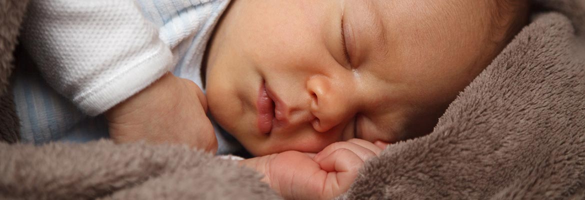
Newborn skin is ill equipped to handle sunlight, temperature stress, penetration of toxic substances and have more water loss. They more readily develop erosions or blisters to trauma of various origins. Learn how to identify troubling spots.
By Emily Jorge, DCNP, FNP-BC
Working with pregnant colleagues shifts our minds to newborns, gift registries, pastel colors, and cute onesies. The skin of the newborn differs from adults in numerous ways. It has less hair, sweat and sebaceous gland secretions; it is thinner and has fewer pigment structures called melanosomes; and it has a decreased number of intercellular attachments. Consequently, newborn skin is ill equipped to handle sunlight, temperature stress, penetration of toxic substances and has more water loss. It more readily develops erosions or blisters to trauma of various origins.
Much of the time growths and rashes on newborn skin can be addressed by another specialty group, but occasionally we in pediatric dermatology encounter a myriad of lesions. Here’s the short-of-the-long of the minor anomalies list:
- Dimpling: This can be found over bony areas, especially above the buttock area or the buttock crease. These are usually of cosmetic consequence only, but can be the first sign of a dysmorphic syndrome—a syndrome where there may be multisystem involvement with malformations. Spinal problems need to be ruled out when deep dimples (typically >5mm in size or further than 2.5cm from the anus) *, sinus tracts, or other lesions such as lipomas, hemangiomas, or hair tufts are present. An ultrasound may suffice within the first three months, but magnetic resonance imaging (MRI) will provide a clear view of the area. This requires sedation of the newborn.
- Sinuses, pits, tags, and cysts: Periauricular sinuses (near the ear), pits, tags, and cyst defects may be unilateral or bilateral and can be associated with facial anomalies or malformations, particularly medial pits of the head and neck. Secondary infections alert the provider to further investigate. Many of these are excised during childhood. The prevalence of preauricular pits and tags is estimated at around .5% to 1.0%. The potential association with these is an increased incidence of hearing or genitourinary defects. This association is decreased in the absence of other syndromic features.* Cysts typically require excision and if they occur midline on the head should prompt imaging. **
- Extra digits: Supernumerary digits are soft tissue duplications without skeletal involvement. Frequently they appear on the ulnar side of the 5th finger and can appear bilaterally or unilaterally. These can be familial and are typically asymptomatic.* Local surgical excision is the treatment.
- Extra nipples: Appearing along a line from the mid underarm to the genitalia, supernumerary nipples develop without areolae, which often results in their misdiagnosis. Investigations demonstrating a relationship between accessory nipples and renal or urogenital anomalies have not be fully elucidated and remain controversial. If physical findings warrant further evaluation, an ultrasound can be considered.
- Umbilical Irritation: Normally the umbilical cord dries up and separates within 1-2 weeks. The surface scars down in the next 1-2 weeks thereafter. However, in some cases granulation tissue may form an umbilical granuloma and rarely secondary infection may occur. Any drainage may warrant further investigation or imaging. Cauterization usually results in the healing of the granuloma.
References:
* Cohen, Bernard A. (2013). Pediatric Dermatology 4th Edition (pp. 23-25). China: Elsevier
** Mancini, Anthony J., & Paller, Amy S. (2016). Hurwitz Clinical Pediatric Dermatology: A Textbook of Skin Disorders of Childhood and Adolescence (5th ed.) (pp. 27-29). Toronto, Canada: Elsevier

